

|
NOTE: image and movie files here are from
authors outside our Institute. For reproduction permissions please
contact the respective authors directly - JYS
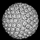
Herpes Simplex Virus-1 A-capsid from 400kV Spot-scan Electron
Cryomicroscopy

Reference
Z. H. Zhou, B.V.V Prasad, J. Jakana, F.R. Rixon, W. Chiu
Baylor College of Medicine,
Journal of Molecular Biology
See updated version and animations from Zhou's page at:
http://hub.med.uth.tmc.edu/~hong/
and
http://ncmi.bioch.bcm.tmc.edu/FacultyLibrary/zhou.html
(c) 1994 Zhou et al. Baylor College of Medicine
EM images with envelope available at:
http://www.uct.ac.za/depts/mmi/stannard/herpes.html
Herpesvirus (entire particle) solved by cryo-electron microscopy and
image reconstruction

 MPEG version (1186K)
 Quicktime version (1382K)
THE ORIGINAL FILE IN IMAGE FORMAT used to be AVAILABLE FROM:
nimh.nih.gov [128.231.98.32 ] as :
pub/nih-image/HerpesVirusMovie.sit.bin
The author of the original movie is
Benes L. Trus, Ph.D.
(E-mail: trus@ipalph.dcrt.nih).
ORIGINAL README FILE
3-D computer reconstruction from cryo-electron micrographs of herpes
simplex virus capsids. Research by scientists at the National Institutes of
Health and the University of Virginia, Charlottesville. The reconstructions
were calculated on DEC-VAX computers using programs supplied by Purdue
University. Reference: "Liquid-Crystalline,Phage-like Packing of
Encapsidated DNA in Herpes Simplex Virus", by F.P.Booy, W.W.Newcomb,
B.L.Trus, J.C.Brown, T.S.Baker, and A.C.Steven, in CELL, Vol 64 pp
1007-1015, March 8, 1991.
Here is the summary of the article cited above -JYS-
SUMMARY
The organization of DNA within the HSV-1 capsid has been
determined by cryoelectron microscopy
and image reconstruction. Purified C-capsids, which are
fully packaged, were compared with A-capsids, which are
empty.
Unlike A-capsids, C-capsids show fine striations and
punctate arrays with a spacing of ~2.6 nm. The packaged
DNA forms a uniformly dense ball, extending radially as far
as the inner surface of the icosahedral (T=16) capsid
shell, whose structure is essentially identical in
A-capsids and in C-capsids. Thus we find no evidence
of the inner T=4 shell previously reported by Schrag et al.
[*SchragJD, Venkataram Prasad BV, Rixon FJ, and Chiu W. (1989).
Three-dimensinal structure of the HSV1 nucleocapsid. Cell
56,651-660)*added by JYS] to be present in C-capsids.
Encapsidated HSV-1 DNA closely resembles that previously visualized
in bacteriophages T4 and _lamda_, thus supporting the idea
of a close parallelism between the respective assembly
pathways of a major family of animal viruses (the
herpesviruses) and a major family of bacterial viruses.
The original file in the Macintosh "Image" format has been translated into a
QuickTimeš movie. The steps were to save the original file from withins Image
as a PICS file, then the PICS file was converted to QuickTimeš with the settings
set to 8 frames per second, 1 key-frame every 8 frames, and the compression
called Apple-Graphics, Grayscale with the Most quality. I tried the JPEG option
and the file size was just 3k more... but the image did not look as sharp.
Published on the Internet. Reproduction granted for educational purposes.
 Go to the
top page Go to the
top page
© 1991 Trus et al.
 |

